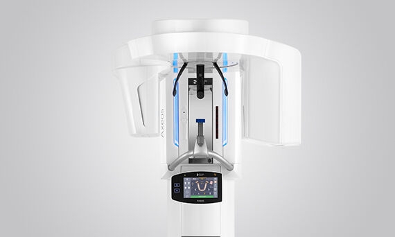
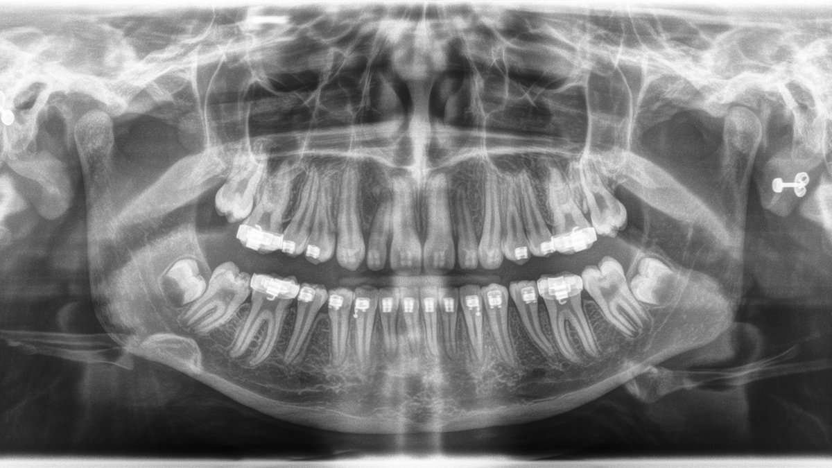
患者ポジショニング
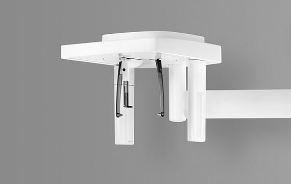
EVIビームライトが、患者さんの位置を示し、電動式の前額面とこめかみサポートが患者さんの頭部を固定。体動による画像のぼやけを防ぎます。
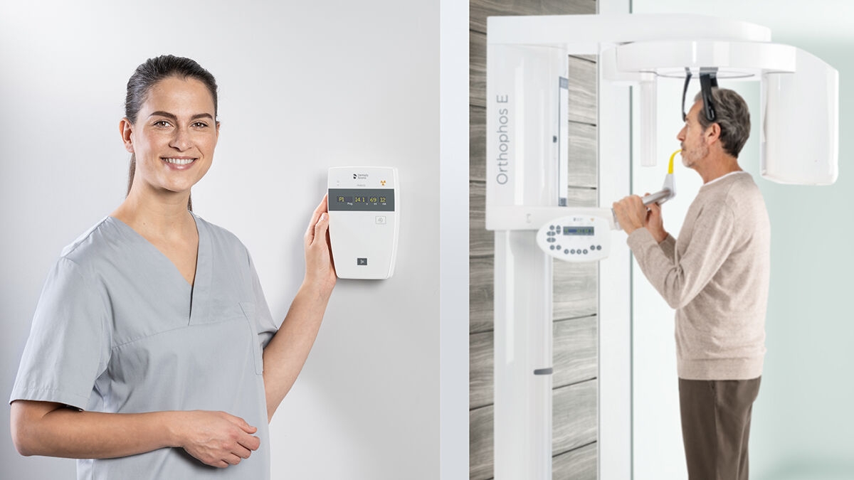
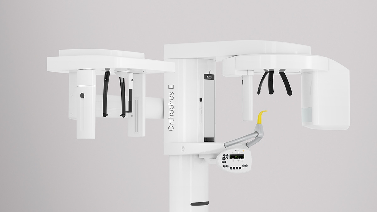
オーソフォスE 2Dは、CsIセンサーテクノロジーとその使いやすいインターフェイスにより、常に信頼性の高い診断を提供します。さらに、 セファロアアームのオプション選択することで、歯科矯正の信頼できるパートナーにもなります。 幅広いサービスであなたの診療を充実させてください。
一般歯科医および歯科矯正医向けのソリューションです。
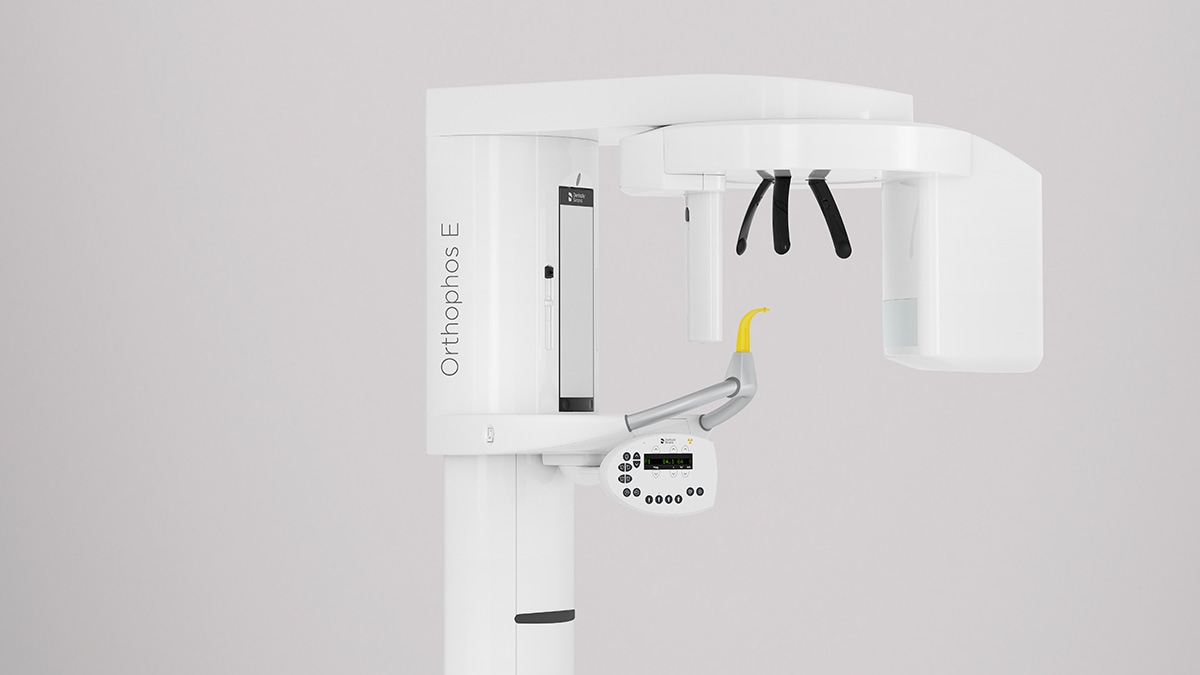
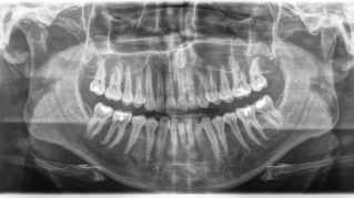
2D CsIセンサーで鮮明な画像が得られます。
電動の3点式ヘッドサポートハンドルが患者さんをしっかり支え、同時にEVIビームライトが撮影範囲に含まれる患者さんの位置を示します。
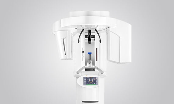
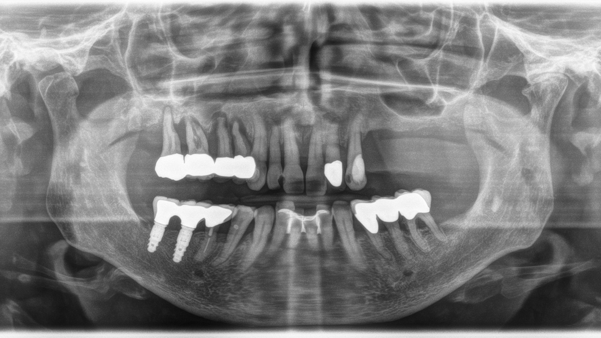
セファロアームは、オプションとして追加することも可能で、また、購入後いつでも後付けすることができます(レフトのみ)。
2Dの基本的な診断のためのプログラム。
コントロールパネルで簡単操作が可能。
Given indications

Given indications
Given indications

Given indications


EVIビームライトが、患者さんの位置を示し、電動式の前額面とこめかみサポートが患者さんの頭部を固定。体動による画像のぼやけを防ぎます。
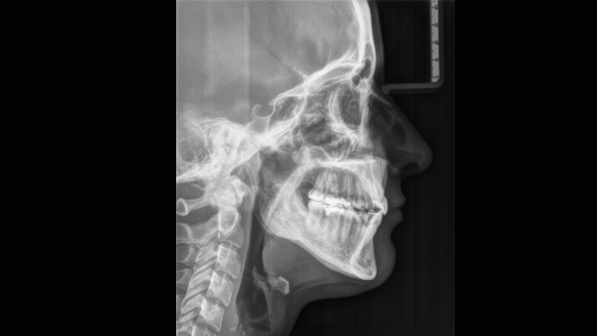
オーソフォスE 2Dには、いつでもセファロアームを取り付けることができます。(レフトのみ)


| Performance features |
Orthophos SL 2D | Orthophos S 2D | Orthophos E |
|---|---|---|---|
| X-ray generator |
60 - 90 kV, 3-16mA | 60 - 90 kV, 3-16mA | 60 - 90 kV, 3-16mA |
| Panoramic exposure time |
P1: max 14,2 s P1 Quickshot: max 9,1 s |
P1: max 14,2 s P1 Quickshot: max 9,1 s |
P1: max 14,2 s |
| Radiation time Ceph |
Standard 9,4 s Quickshot 4,7 s |
Standard 9,4 s Quickshot 4,7 s |
Standard 9,4 s |
| User interface |
EasyPad | EasyPad | MultiPad |
| Patient positioning |
automatic (occlusal bite block) |
automatic (occlusal bite block) |
manual |
| Panorama technology |
DCS | CsI | CsI |
| Autofocus |
yes | yes | - |
| Ceph arm (optional) |
left or right | left or right |
left |
| Ceph unit with 2 sensors |
yes |
yes |
optional |
| Quickshot |
yes |
yes |
- |
| Fields of View |
upgradeable | upgradeable |
- |
| 3D Low Dose |
upgradeable |
upgradeable |
- |
| HD mode |
upgradeable |
upgradeable |
- |
| Base |
optional |
optional |
optional |
| Wheelchair accessible |
yes | yes | yes |
| Remote control |
optional |
optional |
optional |
| Ambient Light | yes | - | - |
| Programme | Orthophos SL 2D | Orthophos S 2D | Orthophos E |
|---|---|---|---|
| Standard panorama image |
P1, P2, P10 |
P1, P2, P10 |
P1, P10 |
| Image detail left side or right side |
P1, P1A, P1C P2, P2A, P2CP10, P10A, P10C BW1 |
P1, P1A, P1C P2, P2A, P2CP10, P10A, P10C BW1 |
P1L, P1R |
| Image detail individual quadrants |
P1, P1A, P1C P2, P2A, P2CP10, P10A, P10C |
P1, P1A, P1C P2, P2A, P2CP10, P10A, P10C |
- |
| Image detail upper or lower jaw |
P1, P1A, P1C P2, P2A, P2CP10, P10A, P10C, P12 |
P1, P1A, P1C P2, P2A, P2CP10, P10A, P10C, P12 |
- |
| Constant magnification |
P1C, P2C, P10C |
P1C, P2C, P10C |
P1C |
| Artifact-reduced |
P1A, P2A, P10A |
P1A, P2A, P10A |
P1A |
| Thick layer front |
P12 |
P12 |
P12 |
| Sinusoidal images |
S1, S3 |
S1, S3 |
S1 |
| Multislice in posterior tooth |
- | - | MS1 |
| Mandibular joint |
TM1.1, TM1.2, TM3 |
TM1.1, TM1.2, TM3 |
TM1.1, TM1.2 |
| Bitewing image |
BW1, BW2 |
BW1, BW2 |
BW1 |
| Ceph (optional) |
C1, C2, C3, C3F, C4 |
C1, C2, C3, C3F, C4 |
C1, C2, C3, C3F, C4 |
はい、デンツプライシロナの3D ImagingはSIDEXIS 4でのみ作動します。しかし、SIDEXIS XGからSIDEXIS 4へのデータの移行は非常に容易です。
オーソフォス SL 2Dおよびオーソフォス S 2Dは3Dアップグレード可能です。 オーソフォス Eはこのオプションを提供していません。